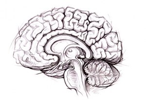
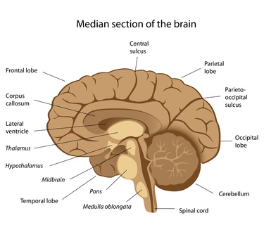
Human normal adult brain
Frozen and paraffin tissue sections, DNA, RNA and proteins from various brain structures
AMYGDALA
The amygdalae (singular: amygdala; also corpus amygdaloideum; Latin, from Greek ?µ??da??, amygdale, 'almond', 'tonsil',listed in the Gray's Anatomy textbook as the nucleus amygdalæ) are almond-shaped groups of nuclei located deep within the medial temporal lobes of the brain in complex vertebrates, including humans. Shown in research to perform a primary role in the processing of memory and emotional reactions, the amygdalae are considered part of the limbic system.
Frozen Tissue Sections
Paraffine Tissue Sections
DNA
Proteins
BASAL GANGLIA
The basal ganglia (or basal nuclei) are a group of nuclei of varied origin in the brains of vertebrates that act as a cohesive functional unit. They are situated at the base of the forebrain and are strongly connected with the cerebral cortex, thalamus and other brain areas. The basal ganglia are associated with a variety of functions, including voluntary motor control, procedural learning relating to routine behaviors or "habits" such as bruxism, eye movements, and cognitive, emotional functions. Currently popular theories implicate the basal ganglia primarily in action selection, that is, the decision of which of several possible behaviors to execute at a given time. Experimental studies show that the basal ganglia exert an inhibitory influence on a number of motor systems, and that a release of this inhibition permits a motor system to become active. The "behavior switching" that takes place within the basal ganglia is influenced by signals from many parts of the brain, including the prefrontal cortex, which plays a key role in executive functions.
Paraffine Tissue Sections
BRAIN ARTERY
The anterior cerebral artery (ACA) is one of a pair of arteries on the brain that supplies oxygenated blood to most medial portions of the frontal lobes and superior medial parietal lobes. The two anterior cerebral arteries arise from the internal carotid artery and are part of the Circle of Willis. The left and right anterior cerebral arteries are connected by the anterior communicating artery.
Paraffine Tissue Sections
BRAIN STEM
In vertebrate anatomy the brainstem (or brain stem) is the posterior part of the brain, adjoining and structurally continuous with the spinal cord. It is usually described as including the medulla oblongata (myelencephalon), pons (part of metencephalon), and midbrain (mesencephalon). Less frequently, parts of the diencephalon are included. The brain stem provides the main motor and sensory innervation to the face and neck via the cranial nerves. Though small, this is an extremely important part of the brain as the nerve connections of the motor and sensory systems from the main part of the brain to the rest of the body pass through the brain stem. This includes the corticospinal tract (motor), the posterior column-medial lemniscus pathway (fine touch, vibration sensation and proprioception) and the spinothalamic tract (pain, temperature, itch and crude touch). The brain stem also plays an important role in the regulation of cardiac and respiratory function. It also regulates the central nervous system, and is pivotal in maintaining consciousness and regulating the sleep cycle. The brain stem has many basic functions including heart rate, breathing, sleeping and eating.
Paraffine Tissue Sections
CEREBELLAR PEDUNCLES
Cerebellar peduncle number six in total may refer to:
Superior cerebellar peduncle - primary output of the cerebellum with mostly fibers carrying information to the midbrain
Middle cerebellar peduncle - carry input fibers from the contralateral cerebral cortex
Inferior cerebellar peduncle - receives proprioceptive information from the ipsilateral side of the body
Paraffine Tissue Sections
cDNA
Proteins
CEREBELLUM
The cerebellum (Latinfor little brain) is a region of the brain that plays an important role in motor control. It may also be involved in some cognitive functions such as attention and language, and in regulating fear and pleasure responses, but its movement-related functions are the most solidly established. The cerebellum does not initiate movement, but it contributes to coordination, precision, and accurate timing. It receives input from sensory systems of the spinal cord and from other parts of the brain, and integrates these inputs to fine tune motor activity. Because of this fine-tuning function, damage to the cerebellum does not cause paralysis, but instead produces disorders in fine movement, equilibrium, posture, and motor learning.
Frozen Tissue Sections
Paraffine Tissue Sections
cDNA
Total RNA
Membran Proteins
Proteins
CEREBRAL CORTEX
The cerebral cortex is a sheet of neural tissue that is outermost to the cerebrum of the mammalian brain. It comprises a major portion of the brain (about two-thirds) and includes the cerebrum and cerebellum. It is the most developed section of the brain and plays a critical role in memory, attention, perceptual awareness, thought, language, and consciousness. It consists of up to six horizontal layers, each with a different composition in terms of neurons and connectivity. The human cerebral cortex is 2–4 mm (0.08–0.16 inches) thick.
Frozen Tissue Sections
Paraffine Tissue Sections
cDNA
Total RNA
Membran Proteins
Proteins
CEREBRAL MENINGES
The meninges is the system of membranes which envelops the central nervous system. In mammals, the meninges consist of three layers: the dura mater, the arachnoid mater, and the pia mater. The primary function of the meninges and of the cerebrospinal fluid is to protect the central nervous system.
Frozen Tissue Sections
Paraffine Tissue Sections
cDNA
Total RNA
Membran Proteins
Proteins
CHOROID PLEXUS
The choroid plexus (from Greek khorion "membrane enclosing the fetus, afterbirth"; "plexus": Mod.L., lit. "braid, network") is a structure in the ventricles of the brain where cerebrospinal fluid (CSF) is produced. The choroid plexus consists of modified ependymal cells.
Paraffine Tissue Sections
DIENCEPHALON
The diencephalon ("interbrain") is the region of the vertebrate neural tube which gives rise to posterior forebrain structures. In development, the forebrain develops from the prosencephalon, the most anterior vesicle of the neural tube which later forms both the diencephalon and the telencephalon. In adults, the Diencephalon appears at the upper end of the brain stem, situated between the cerebrum and the brain stem. It is made up of four distinct components: the thalamus, the subthalamus, the hypothalamus and the epithalamus.
Frozen Tissue Sections
Paraffine Tissue Sections
cDNA
Total RNA
Proteins
HIPPOCAMPUS
The hippocampus is a major component of the brains of humans and other vertebrates. It belongs to the limbic system and plays important roles in the consolidation of information from short-term memory to long-term memory and spatial navigation. Humans and other mammals have two hippocampi, one in each side of the brain. The hippocampus is closely associated with the cerebral cortex, and in primates is located in the medial temporal lobe, underneath the cortical surface. It contains two main interlocking parts: Ammon's horn and the dentate gyrus.
Frozen Tissue Sections
Paraffine Tissue Sections
cDNA
Total RNA
Membran Proteins
Proteins
INSULA
In each hemisphere of the mammalian brain the insular cortex (often called insula, insulary cortex or insular lobe) is a portion of the cerebral cortex folded deep within the lateral sulcus, the fissure separating the temporal and the frontal lobes. The insulae are believed to be involved in consciousness and play a role in diverse functions usually linked to emotion or the regulation of the body's homeostasis. These functions include perception, motor control, self-awareness, cognitive functioning, and interpersonal experience. In relation to these it is involved in psychopathology. The insular cortex is divided into two parts: the larger anterior insula and the smaller posterior insula in which more than a dozen field areas have been identified. The cortical area overlying the insula towards the lateral surface of the brain is the operculum (meaning "lid"). The opercula areformed from parts of the enclosing frontal, temporal and parietal lobes.
Paraffine Tissue Sections
cDNA
Proteins
FRONTAL LOBE
The frontal lobe is an area in the brain of mammals, located at the front of each cerebral hemisphere and positioned anterior to (in front of) the parietal lobe and superior and anterior to the temporal lobes. It is separated from the parietal lobe by a space between tissues called the central sulcus, and from the temporal lobe by a deep fold called the lateral (Sylvian) sulcus. The precentral gyrus, forming the posterior border of the frontal lobe, contains the primary motor cortex, which controls voluntary movements of specific body parts.
Frozen Tissue Sections
Paraffine Tissue Sections
cDNA
Total RNA
Proteins
OCCIPITAL LOBE
The occipital lobe is the visual processing center of the mammalian brain containing most of the anatomical region of the visual cortex. The primary visual cortex is Brodmann area 17, commonly called V1 (visual one). Human V1 is located on the medial side of the occipital lobe within the calcarine sulcus; the full extent of V1 often continues onto the posterior pole of the occipital lobe. V1 is often also called striate cortex because it can be identified by a large stripe of myelin, the Stria of Gennari. Visually driven regions outside V1 are called extrastriate cortex. There are many extrastriate regions, and these are specialized for different visual tasks, such as visuospatial processing, color discrimination and motion perception. The name derives from the overlying occipital bone, which is named from the Latin oc- + caput, "back of the head".
Frozen Tissue Sections
Paraffine Tissue Sections
DNA
Total RNA
Proteins
PARIETAL LOBE
The parietal lobe is a part of the brain positioned above (superior to) the occipital lobe and behind (posterior to) the frontal lobe. The parietal lobe integrates sensory information from different modalities, particularly determining spatial sense and navigation. For example, it comprises somatosensory cortex and the dorsal stream of the visual system. This enables regions of the parietal cortex to map objects perceived visually into body coordinate positions. The name derives from the overlying parietal bone, which is named from the Latin paries-, wall.
Tissue Sections
DNA
Total RNA
Proteins
TEMPORAL LOBE
The temporal lobe is a region of the cerebral cortex that is located beneath the Sylvian fissure on both cerebral hemispheres of the mammalian brain. The temporal lobe is involved in auditory perception and is home to the primary auditory cortex. It is also important for the processing of semantics in both speech and vision. The temporal lobe contains the hippocampus and plays a key role in the formation of long-term memory.
Tissue Sections
DNA
Total RNA
Proteins
MEDULLA OBLONGATA
The medulla oblongata is the lower half of the brainstem. In discussions of neurology and similar contexts where no ambiguity will result, it is often referred to as simply the medulla. The medulla contains the cardiac, respiratory, vomiting and vasomotor centers and deals with autonomic, involuntary functions, such as breathing, heart rate and blood pressure.
Tissue Sections
DNA
Total RNA
Proteins
MIDBRAIN
The midbrain or mesencephalon (from the Greek mesos - middle, and enkephalos - brain) is a portion of the central nervous system associated with vision, hearing, motor control, sleep/wake, arousal (alertness), and temperature regulation. Anatomically, it comprises the tectum (or corpora quadrigemina), tegmentum, the ventricular mesocoelia (or "iter"), and the cerebral peduncles, as well as several nuclei and fasciculi. Caudally the mesencephalon adjoins the pons (metencephalon) and rostrally it adjoins the diencephalon (Thalamus, hypothalamus, etc.). The midbrain is located below the cerebral cortex, and above the hindbrain placing it near the center of the brain.
Tissue Sections
OLFACTORY REGION
The olfactory system is the sensory system used for olfaction, or the sense of smell. Most mammals and reptiles have two distinct parts to their olfactory system: a main olfactory system' and an accessory olfactory system. The main olfactory system detects volatile, airborne substances, while the accessory olfactory system senses fluid-phase stimuli. Behavioral evidence indicates that most often, the stimuli detected by the accessory olfactory system are pheromones. The olfactory system is often spoken of along with the gustatory system as the chemosensory senses because both transduce chemical signals into perception.
Tissue Sections
DNA
Total RNA
Proteins
OPTIC NERVE
The optic nerve, also known as cranial nerve 2, transmits visual information from the retina to the brain. Derived from the embryonic retinal ganglion cell, a diverticulum located in the diencephalon, the optic nerve does not regenerate after transection.
Tissue Sections
DNA
Proteins
PINEAL GLAND
The pineal gland (also called the pineal body, epiphysis cerebri, epiphysis, conarium or the "third eye") is a small endocrine gland in the vertebrate brain. It produces the serotonin derivative melatonin, a hormone that affects the modulation of wake/sleep patterns and seasonal functions. Its shape resembles a tiny pine cone (hence its name), and it is located near the centre of the brain, between the two hemispheres, tucked in a groove where the two rounded thalamic bodies join.
Tissue Sections
PITUITARY
In vertebrate anatomy the pituitary gland, or hypophysis, is an endocrine gland about the size of a pea and weighing 0.5 grams (0.018 oz) in humans. It is not a part of the brain. It is a protrusion off the bottom of the hypothalamus at the base of the brain, and rests in a small, bony cavity (sella turcica) covered by a dural fold (diaphragma sellae). The pituitary is functionally connected to the hypothalamus by the median eminence via a small tube called the infundibular stem (Pituitary stalk). The pituitary fossa, in which the pituitary gland sits, is situated in the sphenoid bone in the middle cranial fossa at the base of the brain. The pituitary gland secretes nine hormones that regulate homeostasis.
Tissue Sections
DNA
Total RNA
Proteins
PONS
The pons (pronounced /'p?nz/) is a structure located on the brain stem, named after the Latin word for "bridge" or the 16th-century Italian anatomist and surgeon Costanzo Varolio (pons Varolii).[1] It is cranial to the medulla oblongata, caudal to the midbrain, and ventral to the cerebellum. In humans and other bipeds, this means it is above the medulla, below the midbrain, and anterior to the cerebellum. This white matter includes tracts that conduct signals from the cerebrum down to the cerebellum and medulla, and tracts that carry the sensory signals up into the thalamus.
Tissue Sections
DNA
Proteins
POSTCENTRAL GYRUS
The lateral postcentral gyrus is a prominent structure in the parietal lobe of the human brain and an important landmark. It is the location of the primary somatosensory cortex, the main sensory receptive area for the sense of touch. Like other sensory areas, there is a map of sensory space in this location, called the sensory homunculus.
Tissue Sections
DNA
Total RNA
Proteins
PRECENTRAL GYRUS
The primary motor cortex is a brain region that in humans is located in the posterior portion of the frontal lobe. It works in association with other motor areas including premotor cortex, the supplementary motor area, posterior parietal cortex, and several subcortical brain regions, to plan and execute movements. Primary motor cortex is defined anatomically as the region of cortex that contains large neurons known as Betz cells. Betz cells, along with other cortical neurons, send long axons down the spinal cord to synapse onto the interneuron circuitry of the spinal cord and also directly onto the alpha motor neurons in the spinal cord which connect to the muscles. The primary motor cortex contains a rough map of the body, with different body parts controlled by partially overlapping regions of cortex arranged from the toe (at the top of the cerebral hemisphere) to mouth (at the bottom) along a fold in the cortex called the central sulcus. Each cerebral hemisphere contains a map that controls mainly the opposite side of the body.
Tissue Sections
DNA
Total RNA
Proteins
THALAMUS
The thalamus (from Greek ???aµ??, "inner chamber")[1] is a midline symmetrical structure within the brains of vertebrates including humans, situated between the cerebral cortex and midbrain. Its function includes relaying sensory and motor signals to the cerebral cortex,[2][3] along with the regulation of consciousness, sleep, and alertness. The thalamus surrounds the third ventricle. It is the main product of the embryonic diencephalon.
Tissue Sections
DNA
Total RNA
Proteins